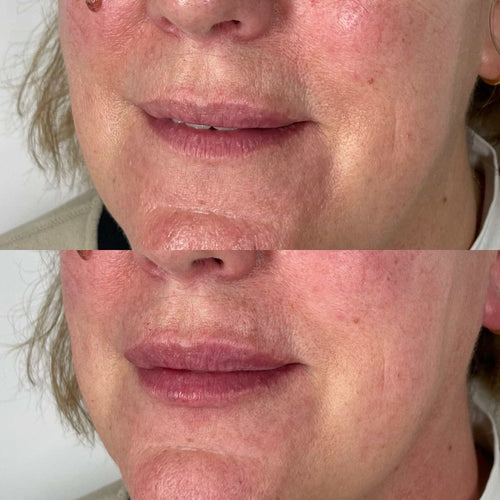
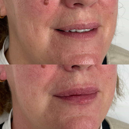
Schedule a Dermal Filler Consultation with Dr. Laura Geige Today
Anatomy of the Preauricular Area
Location
The preauricular area refers to the region of skin located immediately in front of the external ear (pinna).
It’s a triangular-shaped region that sits along the cheek, extending from the corner of the eye to just above the jawline.
Anatomical Structures:
-
Skin: The preauricular area is covered by thin, delicate skin. This skin is more susceptible to irritation and sun damage than other areas of the face.
-
Subcutaneous Tissue: Beneath the skin lies a layer of subcutaneous tissue containing fat, blood vessels, nerves, and lymphatics.
-
Facial Muscles: The preauricular region is innervated by the facial nerve (cranial nerve VII), which controls the muscles responsible for expressions like smiling and frowning.
-
Parotid Gland: This major salivary gland lies partially beneath the preauricular area, contributing to saliva production.

Clinical Significance:
*
Preauricular Sinuses: These are congenital (present at birth) cysts or openings that can occur in the preauricular region. They often present as small, fleshy bumps and may become infected.
*
Lymphadenopathy: Swollen lymph nodes in the preauricular area can be a sign of infection or inflammation in the head, neck, or face.
**Importance:**
Understanding the anatomy of the preauricular area is crucial for medical professionals to accurately diagnose and treat conditions affecting this region.
Skin and Underlying Structures
The preauricular area refers to the region situated immediately anterior to the **external ear (pinna)**, forming a prominent landmark on the face.
Anatomically, this region can be divided into several layers:
Book a Dermal Filler Consultation at It’s Me and You Clinic with Dr. Laura Geige
* **Skin:** The skin of the preauricular area is thin and mobile, characterized by fine wrinkles that become more pronounced with age. It contains a rich network of blood vessels and nerves, supplying sensation to this area.
* **Subcutaneous Tissue (Superficial Fascia):** This layer lies beneath the skin and consists primarily of fat and loose connective tissue. It provides insulation, cushioning, and facilitates movement of the overlying skin.
* **Platysma Muscle:** This sheet-like muscle originates from the fascia covering the upper chest muscles and inserts into the lower jaw, angle of the mouth, and occasionally the preauricular area. When contracted, it tenses the skin of the neck and contributes to facial expressions like frowning and pulling the corners of the mouth downward.
* **Superficial Parotid Lymph Nodes:** The preauricular area is drained by superficial parotid lymph nodes, which are located within the subcutaneous tissue. These nodes filter lymph fluid from the external ear, scalp, face, and part of the oral cavity.
* * **Facial Nerve (CN VII):** The facial nerve branches extensively in the preauricular area, supplying motor innervation to the muscles responsible for facial expression.
* **Retromandibular Vein:** This major vein drains blood from the superficial structures of the face and ear, passing superficially through the preauricular area.
Understanding the anatomy of the preauricular area is crucial in diagnosing and managing various conditions that may affect this region. Common issues include:
* **Preauricular Cysts:** These benign cysts are congenital anomalies filled with fluid or sebaceous material and commonly located near the upper corner of the external ear.

* **Skin Infections:** The preauricular area can be susceptible to bacterial, fungal, or viral infections due to its warm, moist environment and proximity to hair follicles.
* * **Trauma:** Injuries to this region can result from direct blows or lacerations, potentially damaging underlying structures like the facial nerve or lymph nodes.
Muscles
The preauricular area refers to the region situated in front of the ear, specifically on the cheek.
This triangular-shaped region is defined by three key anatomical landmarks: the tragus (a small, cartilaginous projection anterior to the external auditory meatus), the helix (the curved, outer part of the ear), and the corner of the mouth.
The preauricular area contains a unique arrangement of muscles that contribute to facial expressions and movements around the ear.
These muscles are:
– **Levator Labii Superioris:** This muscle originates from the zygomatic bone (cheekbone) and inserts into the upper lip, lifting it up and outward.
It also contributes to elevating the corner of the mouth, thus playing a role in smiling and expressions of happiness.
– **Zygomaticus Major:** This muscle arises from the zygomatic bone and inserts into the corner of the mouth, pulling the lip upward and laterally.
This muscle is primarily responsible for the smile expression, widening the mouth.
– **Zygomaticus Minor:** This muscle shares a similar origin with the zygomaticus major but inserts closer to the upper lip.
It contributes less significantly to smiling and may play a role in raising the upper lip during expressions of amusement or contempt.
– **Risorius:** This muscle originates from the parotid region (the area below and in front of the ear) and inserts into the corner of the mouth.
The risorius is involved in laterally stretching the mouth, contributing to a wide grin or smirk.
Understanding the anatomy and function of these muscles in the preauricular area helps us appreciate the intricate mechanisms underlying facial expressions and communication.
Clinical Significance
Congenital Ear Pits
The preauricular area refers to the region situated anterior and superior to the external ear, often described as in front of, and above, the pinna.
Congenital ear pits are small depressions or dimples found within this preauricular area. They represent a common developmental anomaly, typically appearing bilaterally (on both sides) but occasionally occurring unilaterally.
Clinically, congenital ear pits are generally considered benign and asymptomatic. However, their presence raises certain considerations:
1. **Diagnosis:**
- A detailed physical examination is crucial for identifying ear pits accurately.
- The location, depth, and symmetry of the pits should be meticulously documented.
2.
Potential Complications:
- While rare, congenital ear pits can rarely become infected or obstructed with debris.
- There is a slight increased risk of developing preauricular fistulas, which are abnormal connections between the ear pit and the ear canal or parotid gland.
3.
Aesthetic Concerns:
- Some individuals may find congenital ear pits aesthetically undesirable, leading to concerns about their appearance.
- This can lead to requests for cosmetic procedures such as surgical excision or filling of the pits.
4.
Genetic Associations:
- Congenital ear pits have been linked to certain genetic syndromes, although they are often an isolated finding without other associated anomalies.
- A family history of similar ear pits should be explored during the patient’s evaluation.
Other Conditions Affecting the Area
The preauricular area refers to the region located in front of the ear, specifically the space between the tragus (the small flap of cartilage projecting from the anterior side of the external auditory canal) and the upper portion of the cheek.
Clinical significance within this area is often associated with congenital anomalies.
One notable condition is preauricular sinus or fistula, a developmental defect that results in a small opening or tunnel present just in front of the ear.
This can sometimes lead to infection or inflammation, requiring treatment.
Preauricular pits are also common, appearing as dimples in the skin. They are generally benign and don’t require intervention unless they become irritated or infected.
Other conditions that may affect the preauricular area include:
* **Lymphangioma:** A benign tumor of lymphatic vessels that can present as a swelling, often fleshy or cauliflower-like, in the preauricular region.
* **Nevus:** A birthmark, which can appear as a flat patch of discoloration or a raised bump, and may sometimes be present in the preauricular area.
* **Cutaneous abscesses or infections:** The skin around the ear is prone to infection, which may present as a painful, red swelling.
Schedule Your Dermal Filler Session with Dr. Laura Geige
BeyBey Name Audrey’s JL Alabama Sig Delt Fashionably Balanced Goonie Yoga and Therapy Making Memories London
- Traptox Aka Trapezius Botox Treatment Near Sanderstead, Surrey - January 8, 2025
- Skin Pen Microneedling Near Chiddingfold, Surrey - January 6, 2025
- Skin Pen Microneedling Near Reigate, Surrey - January 4, 2025